
Figure 1.
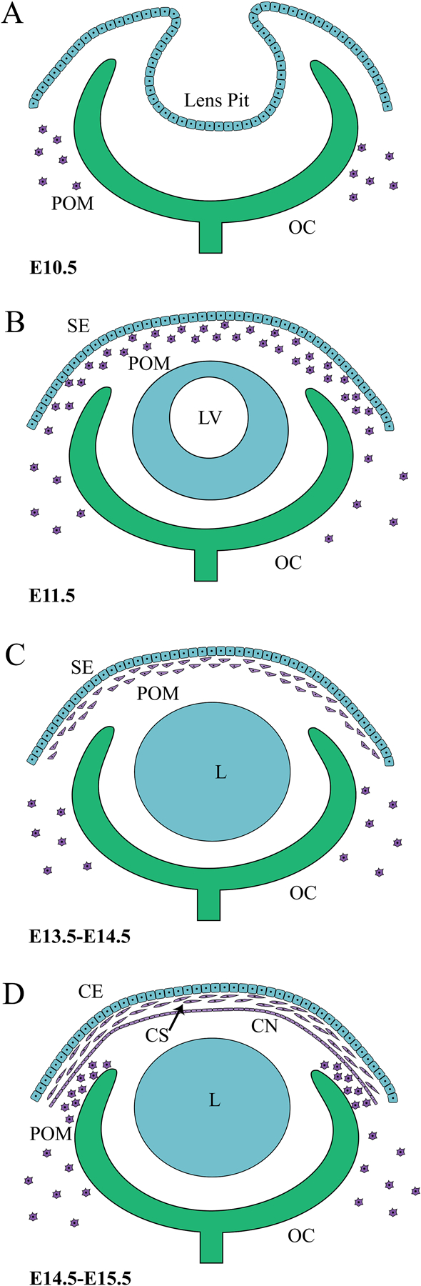
Murine embryonic corneal development and POM migration. (A) The lens pit and optic cup (OC) are formed when the lens placode and optic vesicle invaginate at E10.5. (B) POM cells, derived from the cranial NCCs and mesoderm, migrate at E11.5 into the space between the newly formed surface ectoderm (SE) and lens vesicle (LV). (C) Between E13.5–14.5, while lens fibres close off the lumen of the lens vesicle, the migrated POM cells flatten and condense. (D) By E14.5–15.5, the three layers of the cornea are discernible, with the posterior POM having given rise to the flattened endothelial layer (CN), and outer surface ectoderm developing into the corneal epithelium (CE). The intermediate POM constitutes the corneal stroma (CS) and gives rise to ECM producing keratocytes. Post-E15.5, an additional migration of POM cells along the anterior edge of the optic cup forms the iris stroma and ciliary body stroma. (POM: periocular mesenchyme, OC: optic cup, LV: lens vesicle, SE: surface ectoderm, L: lens, CE: corneal epithelium, CS: corneal stroma, CN: corneal endothelium).