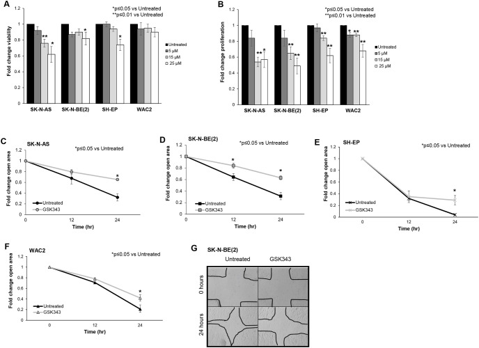
Fig 2. GSK343 decreased neuroblastoma viability, proliferation, and motility.
(A) SK-N-AS, SK-N-BE(2), SH-EP and WAC(2) cells (1.5 × 10³ cells) were treated with increasing concentrations of GSK343 (0, 5, 15, 25 μM) for 24 hours and viability was measured using alamarBlue® assay. GSK343 treatment resulted in decreased viability in SK-N-AS, SK-N-BE(2), and WAC(2) cells. (B) SK-N-AS, SK-N-BE(2), SH-EP and WAC(2) cells (1.5 × 10³ cells) were treated with increasing concentrations of GSK343 (0, 5, 15, 25 μM) for 24 hours and proliferation was measured with using CellTiter® assay. Proliferation was significantly decreased following treatment with GSK343 in all cell lines. (C) SK-N-AS, (D) SK-N-BE(2), (E) SH-EP and (F) WAC(2) cells were treated for 24 hours with GSK343 (15 μM) then plated and allowed to reach 80% confluence. A standard scratch was made in each well and images of the scratch were obtained at 0, 12, and 24 hours. The area of the open wound in pixels was quantified using the ImageJ MRI Wound Healing Tool. By 24 hours, there was a significant decrease in the area of the scratch healed (indicating decreased motility) in cells treated with GSK343 compared to untreated cells. Data were reported as fold change scratch area ± SEM and compared between groups. (G) Representative photomicrographs of SK-N-BE(2) cell wounding assays. GSK343 treated cells (right panel) demonstrated significant reduction in ability to heal the scratch compared to untreated cells (left panel). Data represent at least three biologic replicates.

