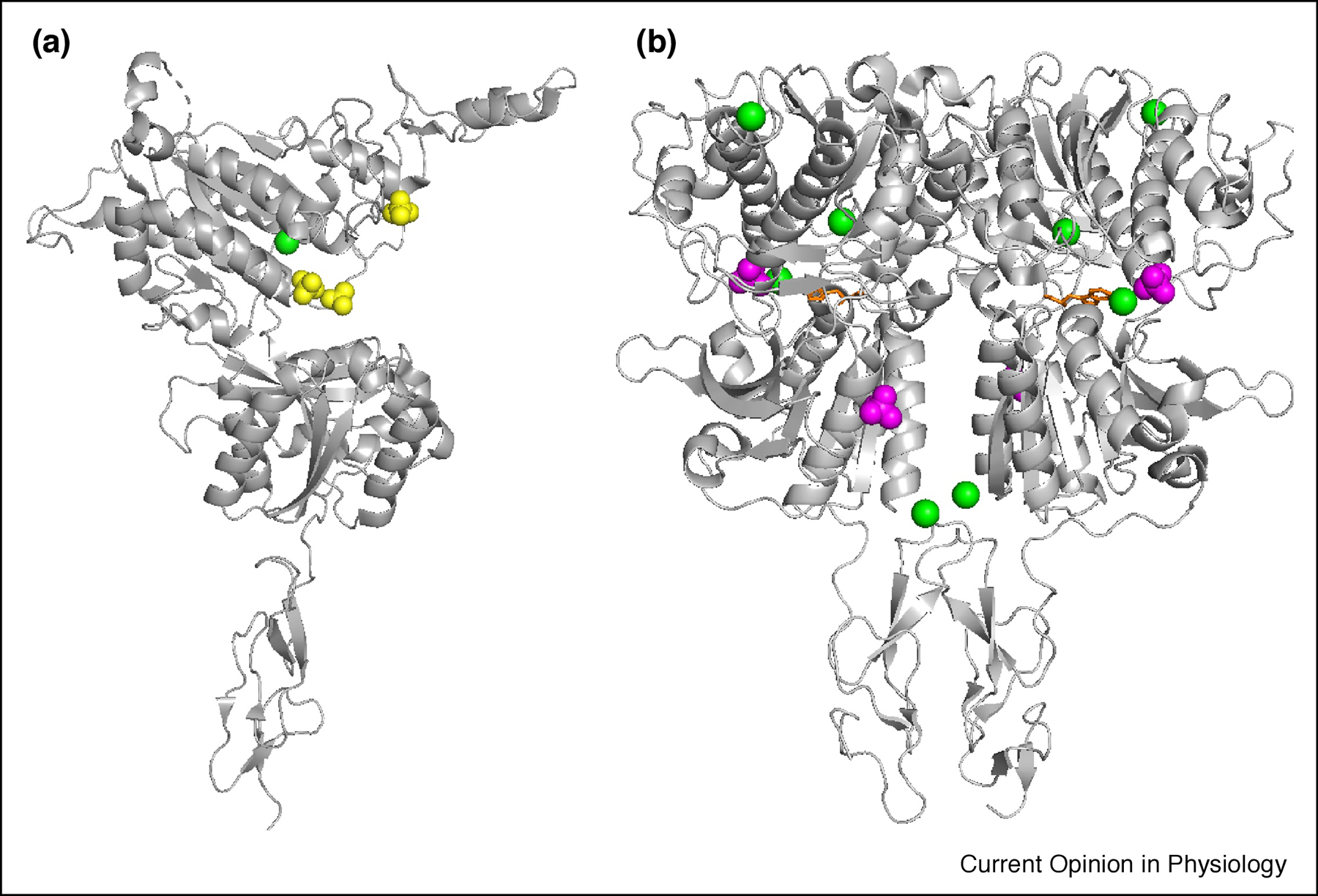
Fig.2.

X-ray Structure of both (A) apo and (B) holo form of CaSR ECD with Cys-rich domain (PDB IDs 5K5T and 5K5S). Green spheres represent Ca2+.SO42− and PO43− are shown in yellow and magenta spheres respectively. L-Trp is indicated by orange sticks.
