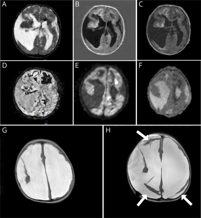
Figure 2. MRI of the Brain.
MRI of 1 day after birth (A–F) and at 10 months (G and H). Postnatal MRI shows porencephalic cysts, bleeding, and ischemic changes. (A) T2. (B) Fluid-attenuated inversion recovery weighted images. (C) T1. (D) Fast field echo showing extensive hypointensive artifacts indicating hemorrhage. (E and F) Diffusion weighted imaging with hyperintensities (E) and corresponding hypointensities apparent diffusion coefficient (F), suggestive of recent ischemia. MRI at 10 months shows extreme hydrocephalus and loss of white matter (G). After receiving a ventriculoperitoneal drain (H), major loss of brain parenchyma is even more obvious; moreover, pericerebral hygromas are now present (white arrows).

