Figure 1.

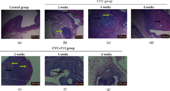
Histopathological changes of the right external jugular vein. HE staining was performed to evaluate the histopathological changes. Representative images are shown. (a) Control group (×10). (b) CVC group at week 2 (×10): vascular intima shedding (black arrow), subintimal hemorrhage (green arrow) and mucoid degeneration (yellow arrow). (c) CVC group at week 4 (×10): granulation tissue organization in venous lumen (black arrow) and inflammatory cell infiltration (yellow arrow). (d) CVC group at week 6 (×20): scar tissue formation under the intima (black arrow). (e) CVC+Y15 group at week 2 (×20: inflammatory cell infiltration (black arrow) and hyaline degeneration (yellow arrow). (f) CVC+Y15 group (×10) at week 4: vascular intima shedding (black arrow). (g) CVC+Y15 group at week 6 (×10). Scale bar: 200 μm.
