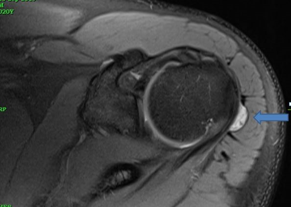
Fig. 1.

An axial proton density fat saturated image showing a well-demarcated hyperintense lesion along the posterior surface of the infraspinatus tendon protruding into the subacromial subdeltoid bursa. The single arrow in axial proton density fat saturated (PDFS) image showing well demarcated hyperintense lesion along the posterior surface of infraspinatus tendon protruding into the subacromial subdeltoid bursa.
