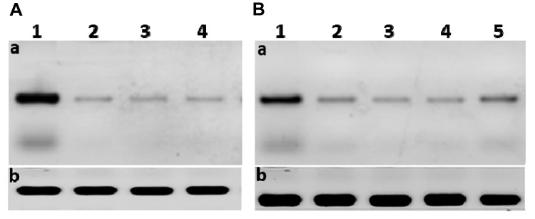
Figure 8.
(A) The scanned densitometry Western blot of viral replication in Huh7 (a) versus β-actin (b); lane 1, protein levels of infected untreated cells: lane 2, infected cells treated with curcumin: lane 3, infected cells treated with CsNPs: lane 4, infected cells treated with curcumin chitosan nanocomposite. (B) The scanned densitometry western blot of viral entry (a) versus β-actin (b) protein levels in positive; lane 1, untreated infected cells: lane 2, cells treated with curcumin: lane 3, cells treated with CsNPs: lane 4, cells treated with curcumin chitosan nanocomposite: lane 5, cells treated with sofosbuvir. HCV core protein at size of 22 KD.

