
Figure 3.
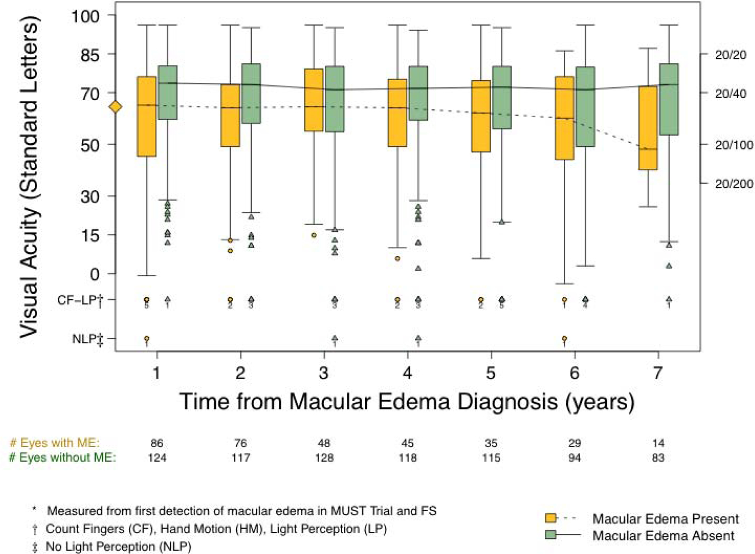
Visual acuity over time among participants with macular edema (ME) stratified by current (time-varying) macular edema status as either absent (green, solid line) or present (orange, dotted line). Macular edema after the initial detection could be either persistent or relapsed. The diamond on the Y-axis denotes median visual acuity at the time of macular edema detection for the entire cohort with macular edema (65 letters; Snellen equivalent 20/50).