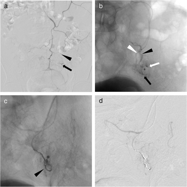
Fig. 3.
A 63-year-old man with sigmoid colonic hemorrhage due to colon cancer. Ultraselective TAE was performed after hemostatic clipping via colonoscopy was unsuccessful. a Inferior mesenteric angiography showed a small and considerably bent vasa recta (black arrowhead) of the sigmoid artery with contrast extravasation (black arrow) at the distal end. b Superselective angiography through the vasa recta (long branch, black arrowhead; short branch, white arrowhead) of the sigmoid artery with a 1.7-F microcatheter (Progreat λ17; Terumo) showed contrast extravasation (black arrow) at the distal end of near clipping (white arrow). After identifying the bleeding site, a 1.7-F microcatheter was inserted into the long branch of the vasa recta as close as possible to the bleeding site (not shown). c Ultraselective angiography through the long branch of the vasa recta showing a microcoil (Galaxy G3 Microcoil; Codman & Shurtleff. Inc.) of 3 mm in diameter and 8 cm long, and two microcoils of 2 mm in diameter and 2 cm long (black arrowhead) placed at the site of contrast extravasation and the bleeding branch. d After ultraselective TAE, contrast extravasation had completely disappeared

