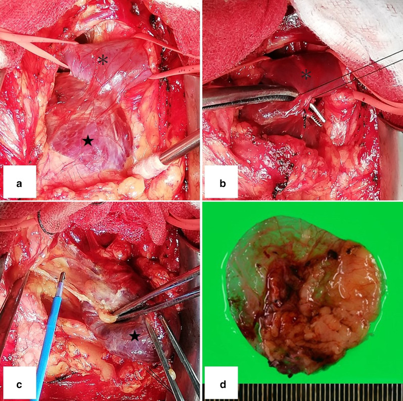
Fig. 2.
The aneurysm (star) was easily dissected from the thymic adipose tissue for the most part, but parts of the aneurysm wall were fragile. The central and peripheral sides of the brachiocephalic vein (asterisk) were secured (a). The brachiocephalic vein (asterisk) and aneurysm (star) were attached by the neck of the aneurysm, which was 8 mm in diameter and was ligated and dissected (b). Aneurysm (star) was not connected to superior vena cava (c). Some parts of the venous walls were thin and fragile (d)

