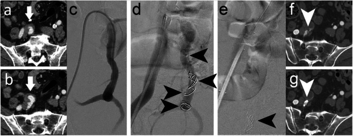
Fig. 2.
Case 1. a, b Preprocedural CTA showing a right common iliac artery aneurysm. c Pre-embolisation DSA imaging of the right IIA (and aneurysm). d 8 mm (distal) and 12 mm diameter (proximal) shape memory polymer plugs were sequentially inserted into the 10 mm diameter right IIA. Arrows indicate the anchor coils and proximal markers of the devices, where the anchor coil is distal to the marker. Shape memory polymer is radiolucent but is between the anchor coil and proximal marker. e Post-embolisation DSA imaging where only the proximal device can be seen in this view (arrow). An iliac stent graft was placed after vessel embolisation. f, g Five-week follow-up CTA showed the vessel was occluded even though a small feeding vessel above the plug (arrow) was perfusing the aneurysm

