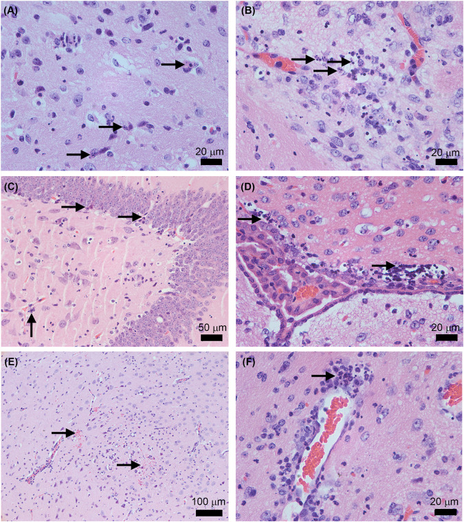
FIGURE 6.
Histopathological features in AG129 mouse brain after the infection with the ZIKV Rio-U1 (Parental) and the Infectious cDNA Clone (IC.RioU1). Karyorrhexis in the cortex of IC.RioU1 (A) and Parental (B) infected animal. Nuclear pyknosis and karyorrhexis in the hippocampus (C) and bordering the choroid plexus (D). Brain hemorrhage foci (E). Perivascular cuffs with a neutrophilic predominance (F). The images are representative of IC.RioU1 (A,C) and Rio-U1 (B,D–F) infection. Black arrows indicate the histopathological features observed in each panel (A–F). The brain sections were stained with hematoxylin and eosin. Data are representative of two independent experiments.

