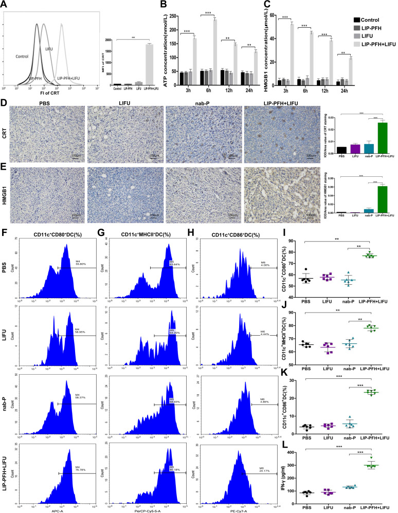
Figure 7.
LIP-PFH nanoparticles triggered by LIFU led to release of DAMPs to promote DC maturation. (A) Membrane exposure of CRT was analyzed by flow cytometry in uptake of LIP-PFH nanoparticles by 4T1 cells at 24h after LIFU stimulating. The mean fluorescence intensity (MFI) was calculated. (B) Extracellular release of ATP from 4T1 cells induced by LIP-PFH nanoparticles triggered by LIFU was measured at 3, 6, 12 and 24h, respectively. (C) HMGB1 released from 4T1 cells induced by LIP-PFH nanoparticles combined with LIFU was measured using ELISA kit, at 3, 6, 12 and 24h, respectively. (D–E) CRT and HMGB1 immunohistochemistry representative images and quantifications of CRT and HMGB1 positive area of tumors (IOD/area value), Scale bar=100μm, n=6; (F–H) representative flow cytometry images of mature DC (including surface markers CD80/MHCII/CD86+DC); (I) the proportion of CD11c+CD80+DC in spleens. (J) The proportion of CD11c+MHCII+DC in spleens; (K) the proportion of CD11c+CD86+DC cells in spleens; (L) the production of IFN-γ in peripheral blood determined by ELISA; n=3, **p<0.01, ***p<0.001.

