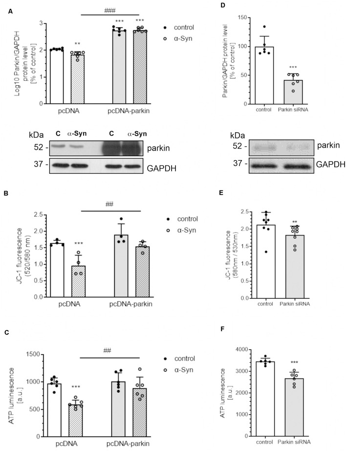
Figure 1.
α-Syn-mediated parkin depletion is responsible for mitochondrial dysfunction in PC12 cells. (A) Parkin immunoreactivity normalized to GAPDH in pcDNA and pcDNA-Parkin PC12 cells treated with α-synuclein (α-Syn) for 24 h at a concentration of 5 μM. Data were log10 transformed and represent the mean value ± SD for 6 independent experiments (n = 6). **p < 0.01, ***p < 0.001 compared to pcDNA control cells; ###p < 0.001 compared to pcDNA cells treated with α-Syn, using two-way ANOVA followed by Bonferroni post hoc test. (B) Mitochondrial membrane potential (ΔΨm) and (C) ATP levels were measured after α-Syn oligomers treatment for 24 h at a concentration of 5 μM in pcDNA and pcDNA-Parkin PC12 cells. Data represent the mean value ± SD (B—n = 4, C—n = 6). ***p < 0.001 compared to pcDNA control cells; ##p < 0.01, compared to pcDNA cells treated with α-Syn, using two-way ANOVA followed by Bonferroni post hoc test. (D) Parkin immunoreactivity normalized to GAPDH in Parkin knock-down PC12 cells. Data were normalized to the corresponding untreated control group (=100%) and represent the mean value ± SEM for six independent experiments (n = 6). ***p < 0.001 compared to corresponding control siRNA, using Student’s t-test. (E) Mitochondrial membrane potential (ΔΨm) and (F) ATP levels were measured in Parkin knock-down PC12 cells. Data represent the mean value ± SD (E—n = 8, F—n = 6). **p < 0.01, ***p < 0.001 compared to corresponding control siRNA, using Student’s t-test.

