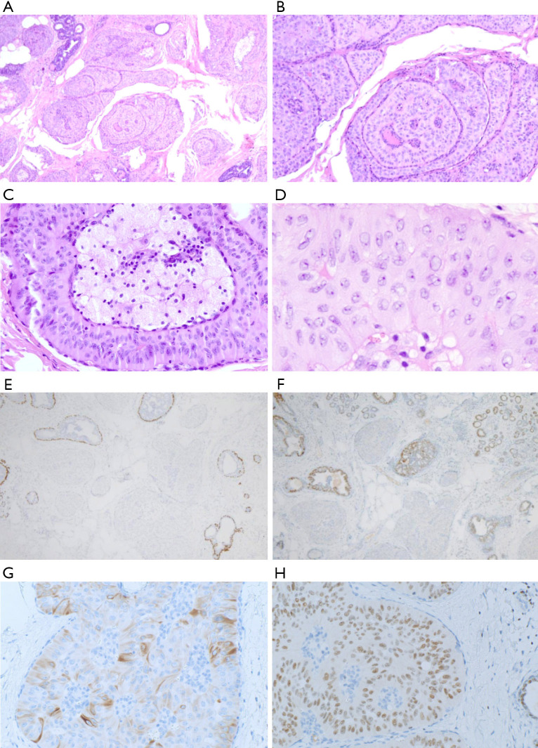
Figure 3.
Histopathological features of tall cell carcinoma of the breast with reverse polarity. (H&E staining; A, ×40; B, ×100) Circumscribed tumor cell nests with fibrovascular cores had infiltrated between normal ducts. Fibrovascular cores contained aggregates of foamy histiocytes. (H&E staining; C, ×200; D, ×400) Nuclei were centralized or located at the top of the cytoplasm (inverted), and mitosis was rare. Nucleus elongation, nuclear clearing, nuclear grooving, and intranuclear pseudo-inclusions were easy to find. (E, ×40) p63 immunostaining showed an absence of myoepithelial cells around and within tumor nests. (F, ×40) Tumor cells were negative for ER staining. (G, ×200) Tumor cells were positive for CK5/6 staining. (H, ×200) Tumor cells were positive for GATA3 staining. ER, estrogen receptor; CK, cytokeratin; GATA3, GATA binding protein 3.

