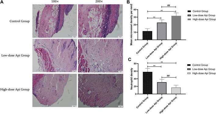
FIGURE 3.
(A) Histopathological features of the flaps were assessed using hematoxylin and eosin (H&E) staining on day 7 after the operation; representative images acquired with a light microscope are shown (magnification,×100, ×200). Api reduced histopathological damage. (B) The microvascular density in zone 2 of the dorsal flap on day 7 after the operation. Api increased microvessel density. (C) Neutrophil density on day 7 after the operation. Api reduced neutrophil density.**p < 0.01 vs. control group. ## p < 0.01 vs. Low-dose Api group.

