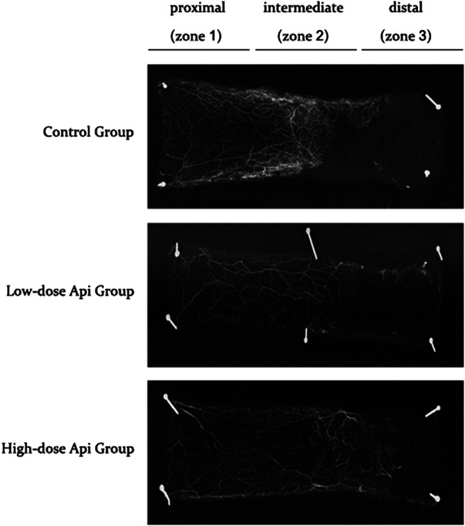
FIGURE 5.

X-ray angiography of the flaps in the control, low-dose Api, and high-dose Api groups on day 7 after the operation. Api improved flap angiographic features.

X-ray angiography of the flaps in the control, low-dose Api, and high-dose Api groups on day 7 after the operation. Api improved flap angiographic features.