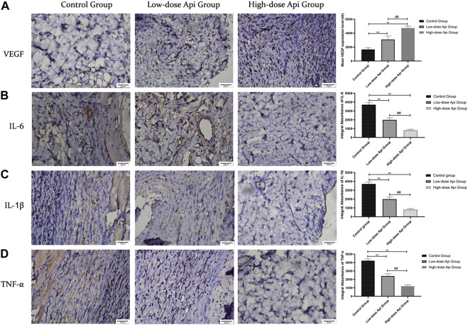
FIGURE 6.
Representative images after immunohistochemical staining for different pro-inflammatory cytokines 7 days after the operation (magnification, ×400). Expression levels of VEGF, TNF-α, IL-1β, and IL-6 in zone 2 of dorsal flaps. Api promoted the expression of VEGF and inhibited the expression of IL-1β, IL-6, and TNF-α. **p < 0.01 vs. control group. ## p < 0.01 vs. Low-dose Api group.

