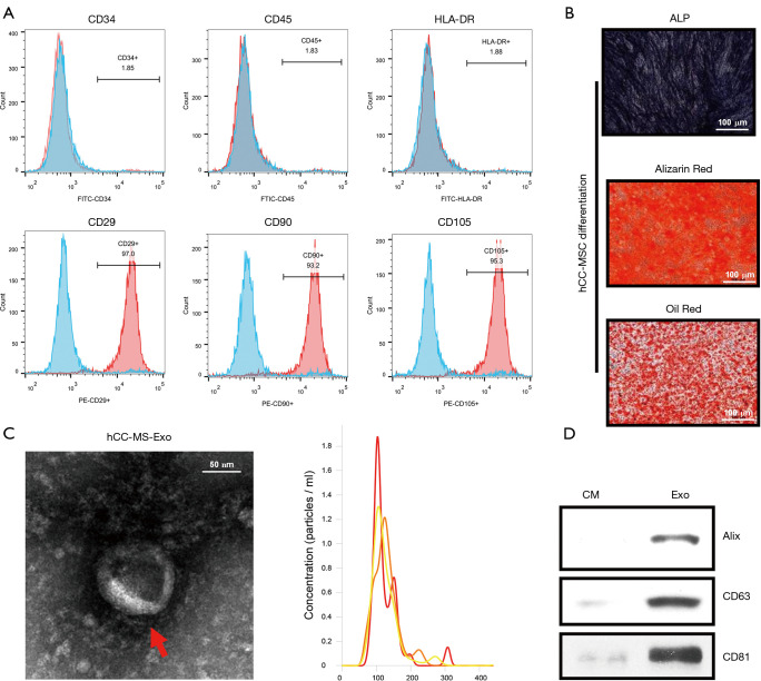
Figure 1.
Characteristics of hCC-MSCs and hCC-MSC-derived exosomes. (A) Flow cytometric analysis of surface markers of hCC-MSCs. (B) Adipogenic and osteogenetic inductions on hCC-MSCs were used for evaluation of the multipotential ability of MSCs to differentiate into cells positively stained with ALP, alizarin red, and oil red O, respectively. (C) Left: transmission electron microscopy images of 2% uranyl acetate-stained exosomes derived from hCC-MSCs (scale bar, 100 nm); right: the sizes of hCC-MSC-derived exosomes (mean: 113 nm). (D) Western blot analysis of Alix, CD63, and CD81 in hCC-MSC-derived exosomes. Exosome-depleted medium as the control. ALP: Alkaline phosphatase; CM: culture medium; Exo: Exosome

