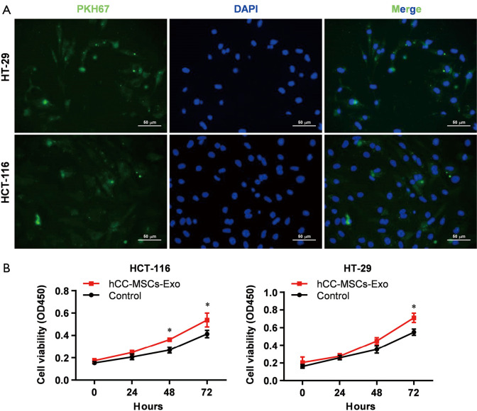
Figure 2.
Uptake of exosomes secreted by hCC-MSCs into HT-29 and HCT-116 cells promoted the cell proliferation. (A) Following fluorescent labeling with cell membrane marker PKH67, hCC-MSC-derived exosomes were incubated with HT-29 and HCT-116 cells. After 24-hour incubation, the cells were subjected to washing and counterstaining with 4',6-diamidino-2-phenylindole (DAPI). Pictures showing the representative results from 3 independent experiments. (B) CCK-8 assay was conducted to study the effect of hCC-MSCs on HT-29 and HCT-116 cell proliferation in vitro. *P<0.05 vs. control group.

