Figure 3.
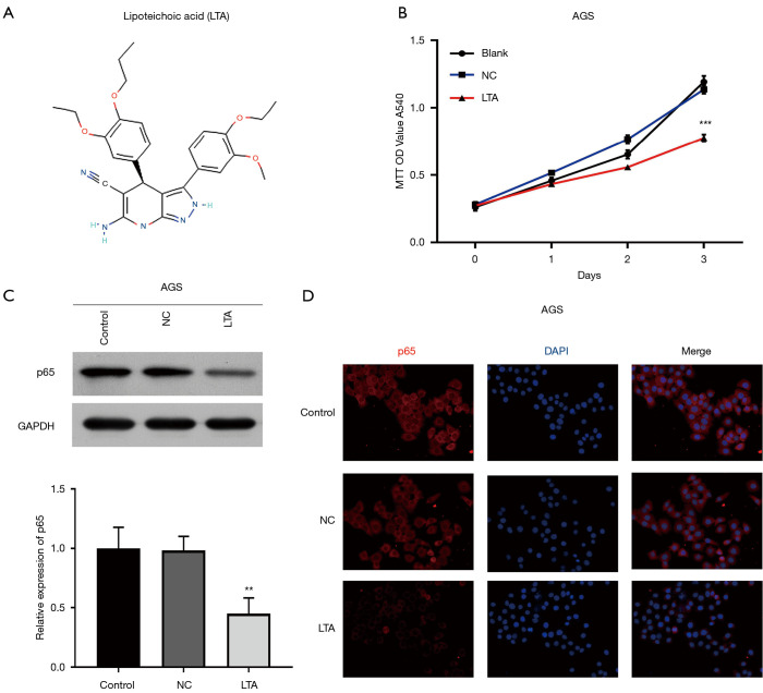
Lipoteichoic acid (LTA) suppressed the proliferation of gastric cancer cells by inhibiting the NF-kappa-B pathway (A) The molecular structure of LTA. (B) AGS cells were treated with LTA (4 µmol/L) for 1, 2 and 3 days. MTT assessed the cell viability. (C) Western blot analyzed the protein level of GAPDH and P65 at 3 days after LTA stimulation. (D) The level of P65 in both the cytoplasm and nucleus of AGS cells was visualized by immunofluorescence assay at 3 days after LTA stimulation. DAPI was used to stain nucleus (blue). All the experiments were carried out in triplicate. **P<0.01, ***P<0.001.

