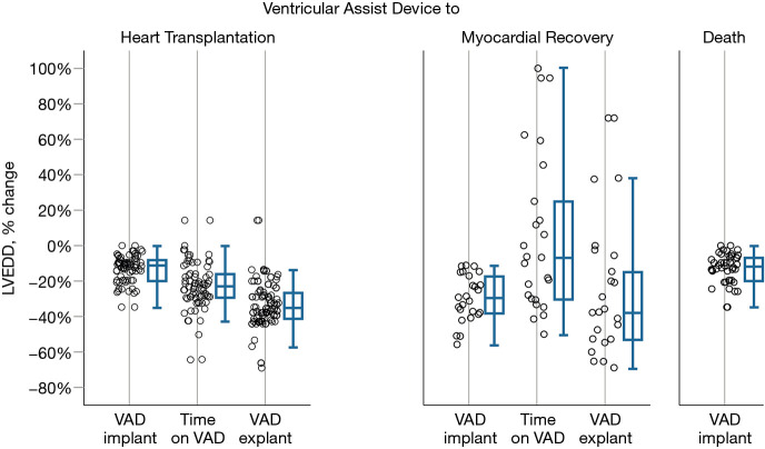
Figure 4.
Percent change in left ventricular end diastolic diameters of three patient groups who had VAD support: those who underwent heart transplantation, recovered and died on VAD. The differences are shown in 3 time points: shortly before VAD implantation, shortly after VAD implantation and after VAD explantation for recovery-patients and after heart transplantation for heart transplant recipients. Patients who recovered had a more pronounced early LVEDD reduction after VAD implantation than those who needed transplantation or those who died. LVEDDs of transplant heart recipients remain smaller, while the LVEDD of patients who had myocardial recovery may either increase or decrease after VAD explantation. The plots show quartiles (boxes represent percentile 25 to 75, the median is the horizontal line in the boxes, whiskers are maximal and minimal values, apart from outliers and extreme values) and individual differences. LVEDD, left ventricular end-diastolic diameter; VAD, ventricular assist device.

