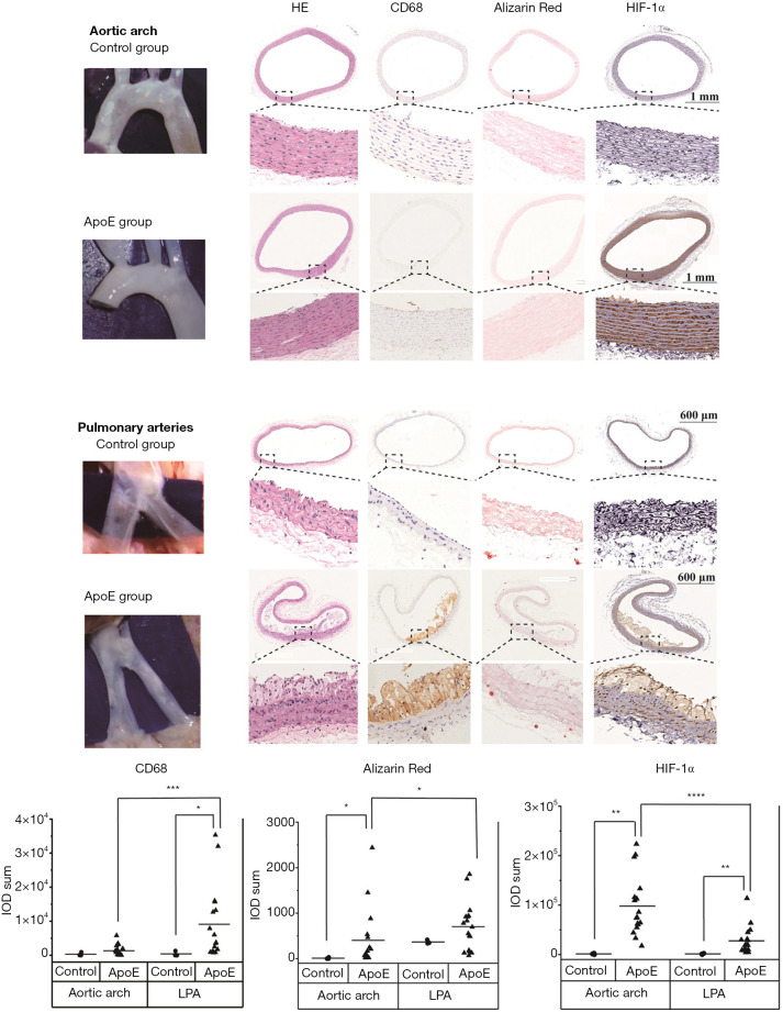
Figure 5.
Pathology and immunohistological study of atherosclerotic lesions in the aortic arch and pulmonary arteries of the normal group (n=3) and the ApoE group (n=17). In the ApoE group, the expression of CD68 and alizarin red was significantly lower in the aortic arch than in the pulmonary arteries. However, the expression of HIF-1α in the aortic arch was significantly higher than that in the pulmonary arteries. The correlations between the groups were calculated by Kruskal-Wallis ANOVA with Dunn’s post-hoc test. *, P≤0.05; **, P≤0.01; ***, P≤0.001; ****, P≤0.0001. LPA, left pulmonary artery; RPA, right pulmonary artery.

