
Figure 5.
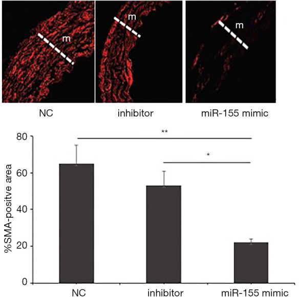
Representative immunofluorescence images of aortas stained with anti-smooth muscle actin (SMA) antibody (red, ×100) in ApoE−/− AS mouse model. Graphs show quantification of VSMC content in the media (m) as either nucleus count or % of SMA-positive area. This method was used to verify that the formed neointima mainly consisted of VSMC and miR-155 mimic group showed mild accumulation in the media while moderate to severe VSMC accumulation was found in the media of miR-155 inhibitor and NC groups. *, miR-155 mimic vs. 155 inhibitor, P<0.01; **, miR-155 mimic vs. NC, P<0.01.