
Fig. 1.
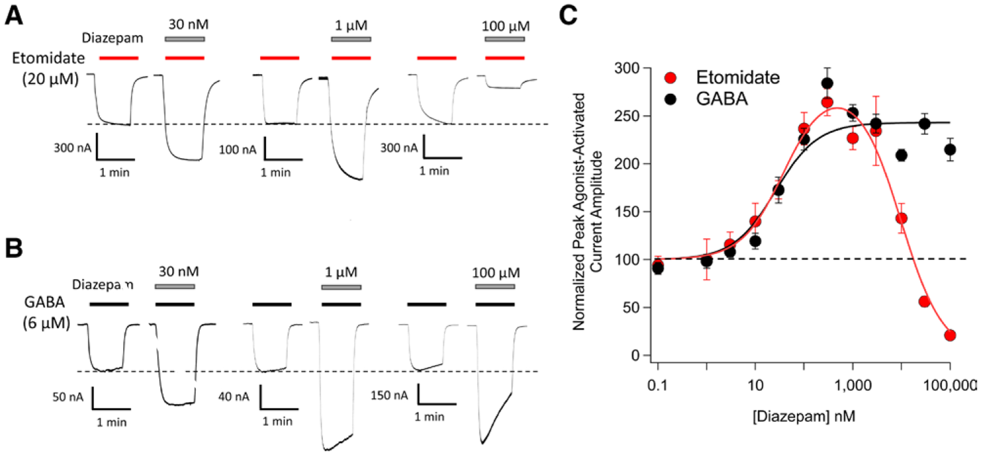
Diazepam modulation of 20 μM etomidate-activated and 6 μM γ-aminobutyric acid (GABA)-activated α1β3γ2L GABAA receptor currents. (A) Representative traces showing the impact of diazepam at the indicated concentrations on etomidate-activated currents. (B) Representative traces showing the impact of diazepam at the indicated concentrations on GABA-activated currents. (C) Diazepam concentration–response curves for etomidate-activated and GABA-activated peak current amplitudes. Each data point represents the mean ± SD derived from five different oocytes. Each curve is a nonlinear least-squares fit of the dataset to either a bell-shaped equation (etomidate-activated currents) or Hill equation (GABA-activated currents). For etomidate-activated peak currents, the diazepam EC50 was 39 nM (95% CI, 27 to 55 nM) and IC50 was 9.6 μM (95% CI, 7.6 to 12 μM). For GABA-activated peak currents, the diazepam EC50 was 26 nM (95% CI, 16 to 41 nM).