
Fig. 2.
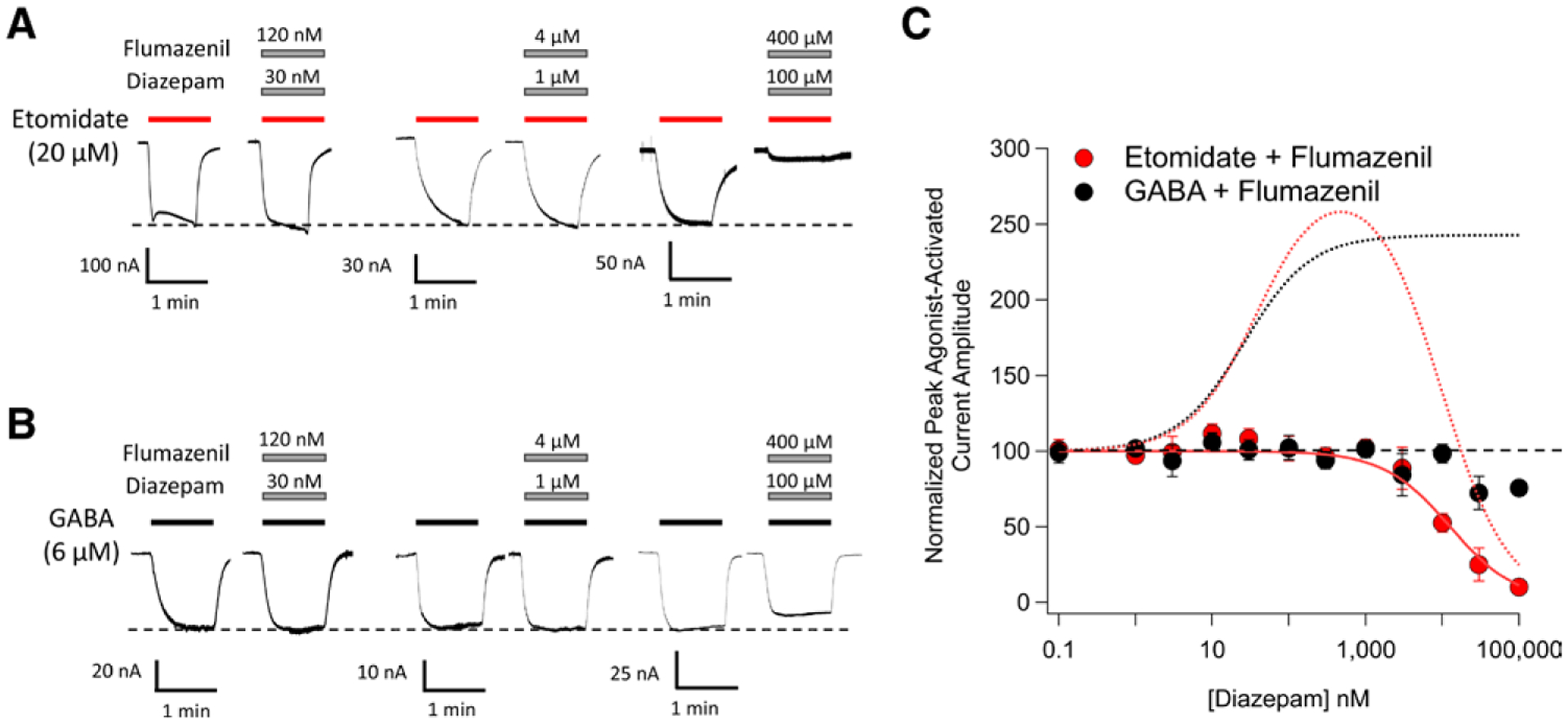
The impact of flumazenil on diazepam modulation of 20 μM etomidate-activated and 6 μM γ-aminobutyric acid (GABA)-activated α1β3γ2L GABAA receptor currents. (A) Representative traces showing the impact of diazepam on etomidate-activated currents in the presence of flumazenil. (B) Representative traces showing the impact of diazepam on GABA-activated currents in the presence of flumazenil. (C) Diazepam concentration–response curves for etomidate-activated and GABA-activated peak current amplitudes in the presence of flumazenil. Each data point represents the mean ± SD derived from five different oocytes. The solid red curve is a fit of the etomidate-activated dataset to a Hill equation yielding a diazepam IC50 of 13 μM (95% CI, 10 to 16 μM). The dotted curves reproduce the fits of the figure 1 data obtained in the absence of flumazenil to illustrate that for both etomidate-activated currents (red dotted curve) and GABA-activated currents (black dotted curve), flumazenil abolishes the nanomolar potentiating action of diazepam. The diazepam:flumazenil concentration ratio was 1:4.