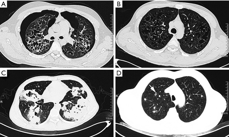
Figure 1.
CT scanning showed that the isolated lung group presented more cystic lesions, including thin-walled cysts accompanied with bilateral pneumothorax (A, case 1), cysts of variable size with small nodules (B, case 2), and thick-walled cysts with multiple masses (C, case 3). Extrapulmonary recidivism (D, case 6) presented poorly defined nodules, with central lucency indicating developing cysts (arrow). CT, computed tomography.

