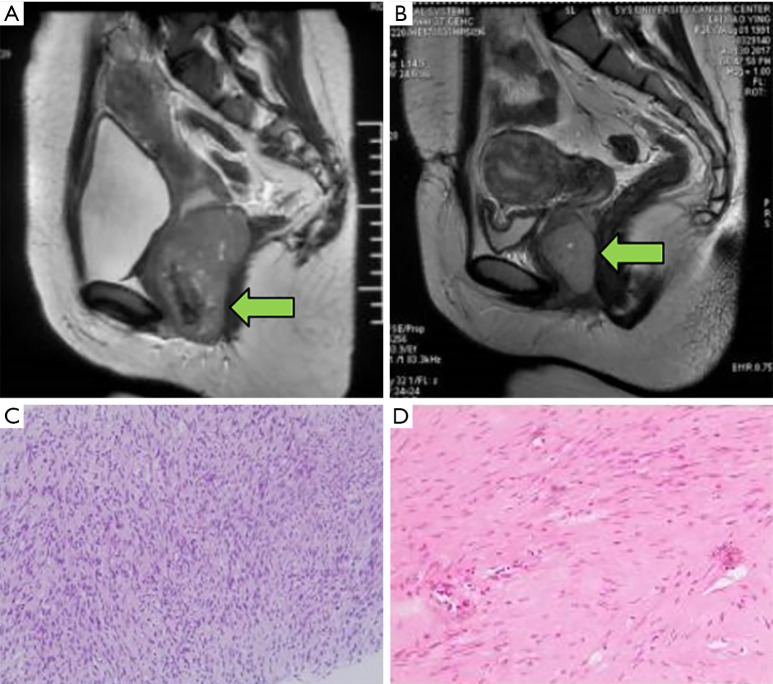
Figure 3.
Comparison of magnetic resonance imaging (MRI) and pathological images pre- and post-neoadjuvant imatinib in a patient with anterior rectal gastrointestinal stromal tumour (GIST) (The “←” points to the location of tumour). (A) Anterior rectal GIST before imatinib (maximum tumour diameter 8.2 cm) and (B) at 10 months after imatinib treatment (maximum tumour diameter 5.0 cm). (C) The pathological image prior to neoadjuvant therapy showing tumour spindle cells (H&E, ×100). (D) The pathological image after imatinib therapy (H&E, ×200).

