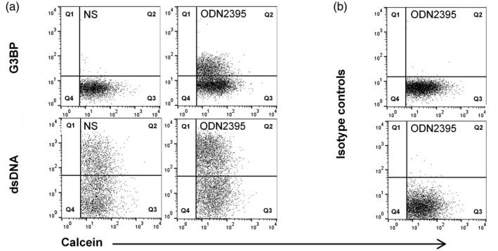
Fig. 2.

Validation of signal‐specificity. Representative fluorescence scatter‐plots from a healthy donor, showing the staining of microvesicles (MVs) from non‐stimulated (NS) and oligodeoxynucleotide (ODN)2395‐stimulated peripheral blood mononuclear cells (PBMCs) with calcein, as a general MV marker, and with (a) anti‐G3BP and anti‐dsDNA antibodies or (b) corresponding isotype controls.
