
Fig. 5.
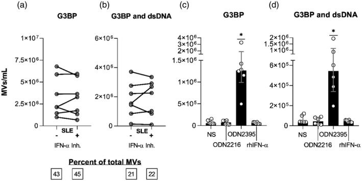
Interferon (IFN)‐α’s role in the Toll‐like receptor (TLR)‐9‐induced release of microvesicles (MVs) from systemic lupus erythematosus (SLE) mononuclear cells. Peripheral blood mononuclear cells (PBMCs) from seven SLE patients [two of whom had active lupus nephritis (LN) at study inclusion] were incubated for 24 h with the TLR‐9 agonist oligodeoxynucleotide (ODN)2395 alone (−) or in combination with the IFN‐α inhibitor IN‐1 (+). MVs released into the culture supernatants were subsequently isolated by ultracentrifugation, quantified and characterized with respect to expression of G3BP and dsDNA by flow cytometry. (a) Concentrations of G3BP‐expressing MVs in toto and (b) G3BP and dsDNA double‐positive MVs in the culture supernatants. Median proportions of MVs bearing these markers are inserted in the boxes below. (c,d) PBMCs from six healthy donors were left non‐stimulated (NS) or were incubated for 24 h with the TLR‐9‐agonist ODN2216, a weak B cell activator and strong IFN‐α inducer; with the TLR‐9 agonist ODN2395, a potent B cell activator and strong IFN‐α inducer; or with recombinant human (rh)IFN‐α. (c) The concentrations of G3BP‐expressing MVs in toto and (d) MVs double‐positive for G3BP and dsDNA in the culture supernatants from non‐stimulated (NS) and stimulated cells. Columns and error bars represent median values and interquartile range. Asterisks above horizontal lines represent P‐values for comparisons between non‐stimulated and stimulated samples. *P < 0·05.