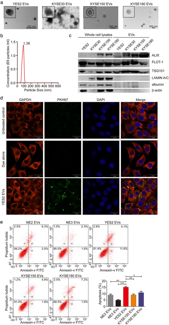
FIGURE 2.

Identification and characterization of EVs derived from ESCC. (a) Representative transmission electron microscopy image of ESCC cell line‐derived EVs (YES2, KYSE30, KYSE150 and KYSE180). (b) Nanoparticle tracking analysis (NTA) indicated the size distribution and concentration of EVs secreted by YES2 cells. (c) Western blot showed expression of EV markers (ALIX, FLOT‐1, and TSG101) in the whole cell lysates and EVs derived from YES2, KYSE30, KYSE150 and KYSE180 cells. LAMIN A/C, albumin and β‐actin were used as non‐exosomal markers. The amount of EVs and cells loaded was normalized to the amount of total proteins. (d) Confocal microscopy images of the internalization of YES2 EVs labelled with 2 nM PKH67 dye in C2C12 myoblasts. Dye alone group was used as a positive control. Untreated control group was used as a negative control. Scale bars, 30 μm. (e) For apoptosis analysis, C2C12 myoblasts were treated with NE2, NE3, YES2, KYSE150 and KYSE180 EVs (10 μg) for 24 h. The cells were collected and stained with annexin V and PI
