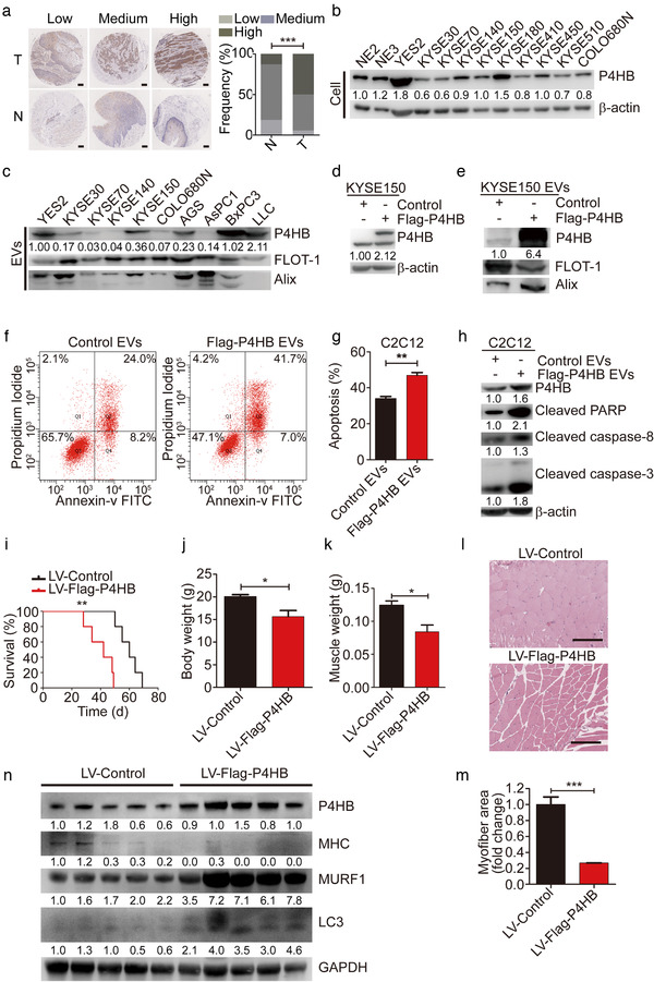
FIGURE 3.

P4HB is identified as a crucial mediator of muscle wasting in vitro and in vivo. (a) Micrographs after immunohistochemistry against P4HB in ESCC tissues (T) and matched adjacent normal tissues (N). Scale bars, 200 μm. The frequency of P4HB expression levels in 77 pairs of human ESCC tissues and matched adjacent normal tissues. (b) The P4HB protein levels in two immortalized esophageal epithelial cell lines (NE2 and NE3) and 10 ESCC cell lines were analyzed by immunoblotting. (c) The P4HB levels in EVs released from ESCC cell lines and other types of cancer cells known as cachexia‐induced cancer cells (AGS, AsPC1, BxPC3, LLC). (d) Overexpressed P4HB protein levels after transfecting the Flag‐P4HB plasmid in KYSE150 cell lines. (e) The P4HB levels in EVs secreted by KYSE150 transfected with Flag‐P4HB plasmid. (f and g) For apoptosis analysis, C2C12 myoblasts were treated with EVs (10 μg) for 24 h derived from KYSE150 cells transfected with Flag‐P4HB plasmid induced by 35 μM cisplatin. (h) Western blot analysis of apoptotic markers in C2C12 myoblasts treated with EVs (10 μg) for 24 h derived from KYSE150 cells transfected with Flag‐P4HB plasmid in combination with 35 μM cisplatin. (i) Kaplan–Meier survival curves of nude mice subcutaneously implanted with KYSE150 cells with stable P4HB overexpression (n = 5), relative to P4HB negative control mice (n = 5). (j and k) Body weight and GA muscle weight changes in KYSE150 implanted mice (n = 5 for each group). (l) Representative micrographs of H&E histology of GA muscle in KYSE150 implanted mice (n = 5 for each group). Scale bars, 150 μm. (m) Quantification of the average myofiber cross‐sectional areas of KYSE150 implanted mice (n = 5 for each group). (n) Western blotting in vivo of MHC, MURF1 and LC3 protein in GA muscle from mice implanted with KYSE150 cells with stable P4HB overexpression (n = 5), relative to P4HB negative control mice (n = 5)
