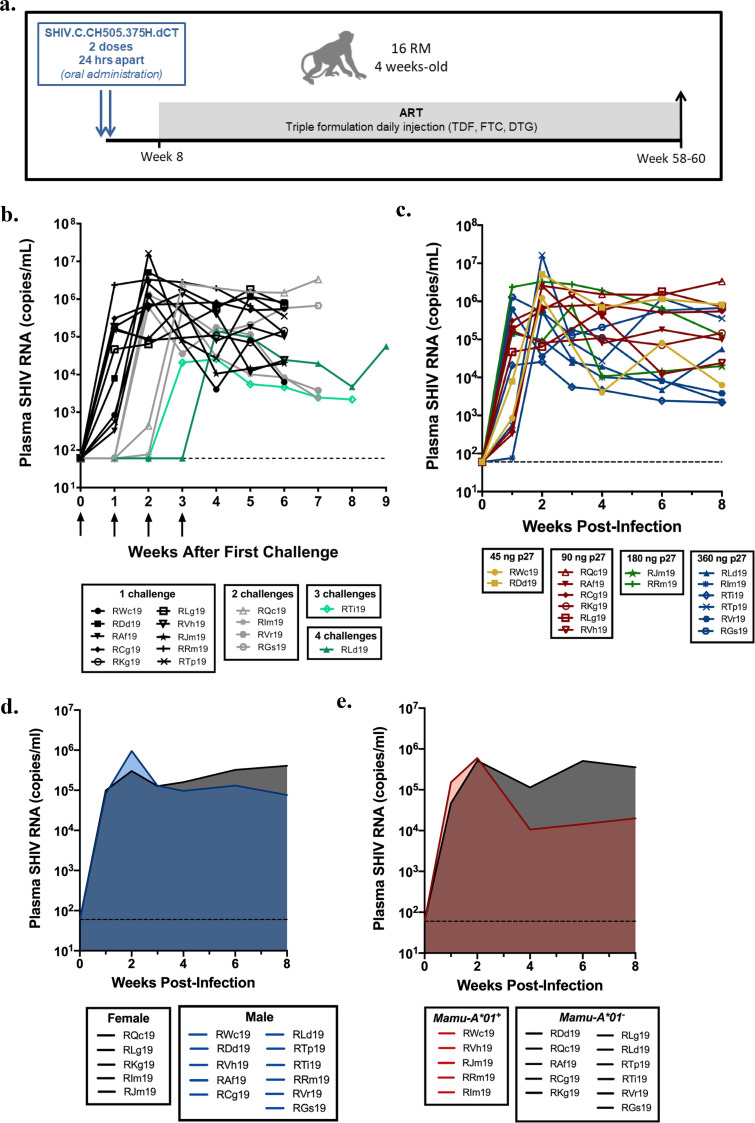
FIG 1.
Infant rhesus macaque model of oral SHIV.C.CH505 infection. (a) Study schematic showing timing and duration of ART. (b to e) Viral replication kinetics prior to ART assessed by number of challenges (b), challenge dose (c), sex (d), and Mamu-A*01 status (e). In panel b, arrows indicate challenge. Plasma viral loads were determined by real-time RT-PCR (n = 16). In panels b and c, each curve represents one animal. Shaded regions in panels d and e represent the median.

