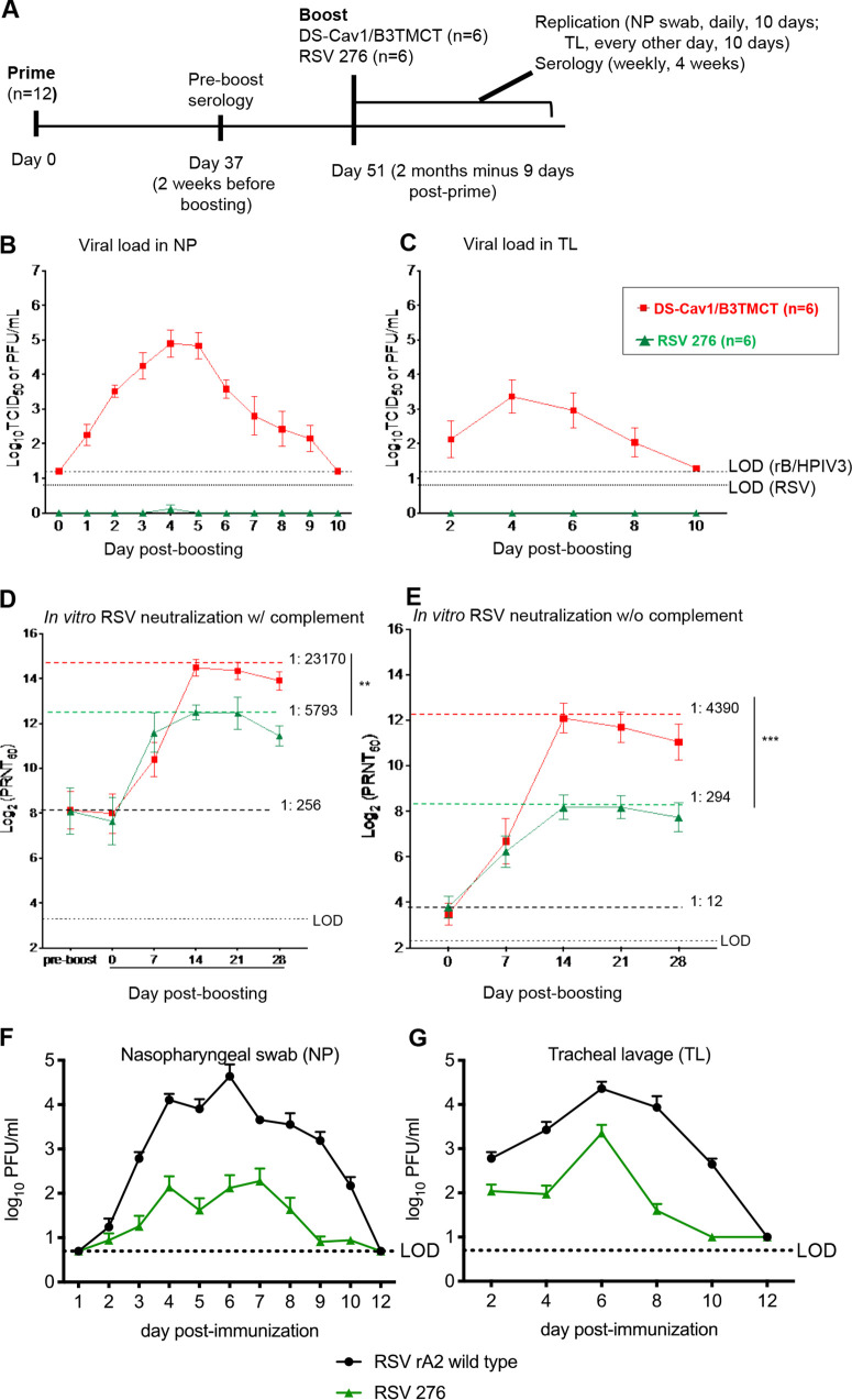
FIG 2.
AGM experiment 1: viral replication and serum RSV-PRNTs in AGMs when the interval between priming and boosting was ∼2 months (2 months minus 9 days). (A) Study design. Twelve AGMs were previously administered a primary infection with one of three RSVs (Table 1) by the combined i.n. and i.t. routes. Sera were collected on day 37 (2 weeks before boosting), and RSV-PRNTs were measured in the presence of complement. The AGMs were organized into two groups of 6 animals each that were balanced with regard to the day 37 RSV-PRNTs, the identity of the priming virus, and the sex ratio (Table 1). On day 51 (2 months minus 9 days) following priming, the groups were boosted with RSV 276 or DS-Cav1/B3TMCT vector by the combined i.n. and i.t. routes. Virus shedding was monitored on days 1 to 10 post-boost with NP and TL samples and virus titration. Sera were collected on days 7, 14, 21, and 28 post-boosting. (B and C) Viral titers in the NP (B) and TL (C) samples shown as means, with brackets indicating standard errors of means (SEMs), and LODs shown as dashed lines (vectors) and dotted lines (RSV). (D) Serum RSV-PRNTs at day 37 postpriming and days 0, 7, 14, 21, and 28 post-boosting, assayed in the presence of complement. (E) Serum RSV-PRNT at days 0, 7, 14, 21, and 28 post-boosting, assayed without complement. Panels D and E are annotated to show the mean serum RSV-PRNTs for the combined two groups at the time of boosting (black dashed lines, with mean arithmetic values shown); in addition, dashed colored lines indicate the highest mean serum RSV-PRNT for each group, with the arithmetic values shown. Mean serum RSV-PRNTs are shown with brackets indicating SEMs. Peak mean titers of two groups were compared by Student's t test. **, 0.001 < P < 0.01; ***, 0.0001 < P < 0.001. (F and G) Replication of RSV 276 and wt rRSV in RSV-seronegative AGMs. RSV-seronegative AGMs were infected by the combined i.n. and i.t. routes with 106 PFU of RSV 276 (n = 8) or wt rRSV (n = 4) in a 1-ml inoculum per site. NP (F) and TL (G) samples were collected daily and every second day, respectively, for 10 days and on day 12. Viral titers were determined by immunoplaque assay and are shown as group means for each time point. Brackets indicate the SEMs, and the LODs are shown as dotted lines. The 8 animals infected with RSV 276 here are the ones shown in Tables 2 and 3 that were subsequently boosted in AGM experiments 2 and 3.

