Figure 4.
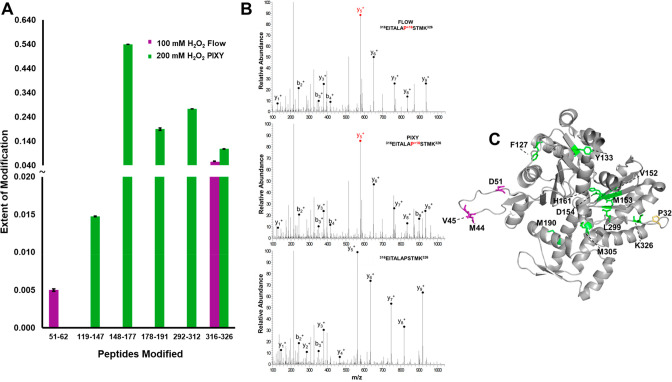
Localization of IC-FPOP modifications. (A) Oxidatively modified peptides within G-actin from the flow system (purple) vs PIXY (green). (B) Tandem MS spectra of modified and unmodified G-actin peptide 316–326 showing b- and y-ions and a +16 FPOP modification on proline in both systems. The MS spectrum of the unmodified peptide is shown at the bottom. (C) FPOP modified residues of G-actin (PDB: 6ZXJ, chain A) are represented in stick representation: 11 modified residues in PIXY (green), 3 modified residues in flow (purple), 1 overlapping modified residue (yellow).

