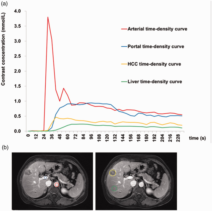
Figure 2.
Dynamic contrast-enhanced magnetic resonance imaging of a 51-year-old male patient with a 10-year history of chronic hepatitis B. Segment VII of liver with pathologically confirmed hepatocellular carcinoma (HCC), grade III. (a) Time-intensity curve of hepatic artery, portal vein, HCC, and liver parenchyma enhancement. (b) Regions of interest: red circle, hepatic artery; blue circle, portal vein; orange polygon, HCC; green circle, liver parenchyma.

