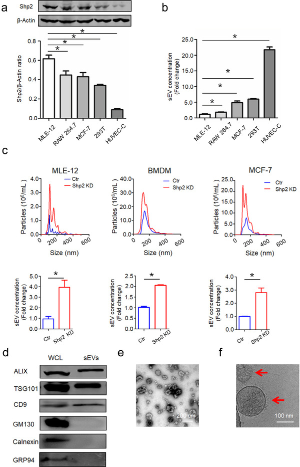
FIGURE 1.

Phosphatase Shp2 regulates secretion of small extracellular vesicles in vitro. [(a) Relative protein level of Shp2 and (b) sEV concentration in different cell lines. sEV concentration represents the quantity of sEVs released by same number of cells. Fold change is compared to MLE‐12 cells. (c) NanoSight quantification of sEV numbers in control and Shp2 KD cells. sEV concentration represents the quantity of sEVs released by same number of cells. Fold change is compared to control. (d) Western blot analysis of sEVs purified from cell culture supernatants from MLE‐12 cells. Whole cell lysates (WCL) and sEVs were blotted for EV‐markers ALIX, TSG101, CD9, and negative control GM130, Calnexin and GRP94.(e)Negatively stained TEM image and (f) Cryo‐EM image (indicated by arrowheads) show purified sEVs from MLE‐12 cells. Data from three independent experiments are shown. *P < 0.05, **P < 0.01, and ***P < 0.001]
