
FIGURE 2.
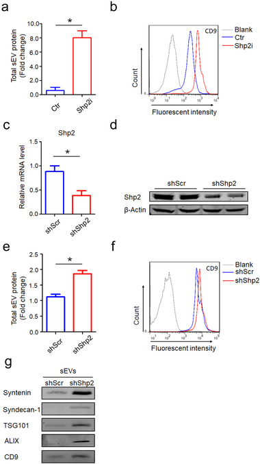
Pharmacologic inhibition and shRNA‐mediated down‐regulation of Shp2 increases sEV secretion of epithelial cells. [(a) Quantification of protein levels in the sEVs obtained from Ctr (DMSO) and Shp2i (PHPS1 20 μM, Shp2i is short for Shp2 inhibition) treated cells (MLE‐12 cells). Fold change is compared to Ctr. (b) After being absorbed on latex beads, sEVs obtained from Ctr (DMSO) and Shp2i (PHPS1 20 μM) treated cells (MLE‐12 cells) were analyzed by flow cytometry with the CD9 antibody.(c) Efficiency of Shp2 depletion in Shp2 KD stable epithelial cell lines (MLE‐12 cells). (d) Western blot analysis of Shp2 in Shp2 KD stable epithelial cell lines (MLE‐12 cells). (e) Quantification of protein levels in the sEVs obtained from shScr and shShp2 stable epithelial cell lines (MLE‐12 cells). Fold change is compared to shScr. (f) After being absorbed on latex beads, sEVs obtained from shScr and shShp2 stable epithelial cell lines (MLE‐12 cells) were analyzed by flow cytometry with the CD9 antibody. (g) Western blot analysis of sEVs purified from cell culture supernatants from equal numbers of shScr and shShp2 stable epithelial cell lines (MLE‐12 cells). sEVs were blotted for Syntenin, Syndecan‐1, TSG101, ALIX and CD9. Data from three independent experiments are shown. *P < 0.05, **P < 0.01, and ***P < 0.001]