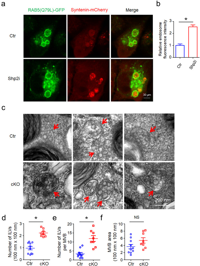
FIGURE 3.

Increases in number of intracellular vesicles (ILVs) by Shp2 inhibition or depletion. [(a) Confocal micrographs show the accumulation of Syntenin‐mCherry inside the lumen of enlarged endosomes outlined by RAB5(Q79L)‐GFP in Ctr (DMSO) and Shp2i (SHP099 20 μM) treated cells (MCF‐7). (b) Relative fluorescence intensity of Syntenin‐mCherry in RAB5(Q79L)‐GFP endosomes indicates relative ILV numbers. Each quantification was performed considering at least 30–50 RAB5(Q79L) endosomes. (c) Representative TEM images of ATII cells in Ctr and cKO (ATII conditional Shp2 KO) mice. Red arrows indicate MVBs containing typical intraluminal vesicles (ILVs). (d) The ILV density in MVBs. (e) The number of ILVs per MVB. (f) Quantitative analysis of MVB area. The ILV density, number of ILVs per MVB and MVB area in 8–10 profiles of different ATII cells were counted and only MVBs containing typical ILVs were counted. Data from three independent experiments are shown. *P < 0.05, **P < 0.01, and ***P < 0.001. NS, non significant]
