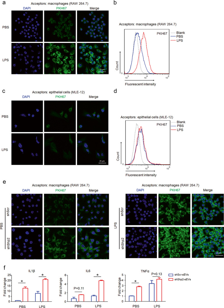
FIGURE 6.

Epithelial Shp2 stable KD promotes sEVs transferring from epithelial cells to macrophages and facilitates macrophage activation in vitro. [(a) Confocal images show uptake of epithelial sEVs by macrophages. PKH 67‐labeled epithelial cells (MLE‐12 cells) were seeded into the upper chamber of the transwell insert. Macrophages (RAW 264.7) were seeded in the lower chamber. After 24 h co‐culture with PBS or LPS (100 ng/ml), macrophages were analyzed by confocal microscopy. (b) Flow cytometry analysis of macrophages in the lower chamber. (c) Confocal images show uptake of macrophage sEVs by epithelial cells. PKH 67‐labeled Macrophages (RAW 264.7) were seeded into the upper chamber of the transwell insert. Epithelial cells (MLE‐12 cells) were seeded in the lower chamber. After 24 h co‐culture with PBS or LPS (100 ng/ml), epithelial cells were analyzed by confocal microscopy. (d) Flow cytometry analysis of epithelial cells in the lower chamber. (e) Confocal images show uptake of epithelial sEVs by macrophages. PKH 67‐labeled shScr and shShp2 stable epithelial cells (MLE‐12 cells) were seeded into the upper chamber of the transwell insert. Macrophages (RAW 264.7) were seeded in the lower chamber. After 24 h co‐culture with PBS or LPS (100 ng/ml), macrophages were analyzed by confocal microscopy. (f) Macrophages (RAW 264.7) were treated with sEVs derived from shScr and shShp2 stable epithelial cell lines (MLE‐12 cells). After 4 h, cells were stimulated with PBS or LPS (100 ng/ml) for 2 h. The mRNA levels of inflammatory cytokine IL1β, IL6 and TNFα were determined by qPCR. Fold change is compared to shScr‐sEVs in PBS. Data from three independent experiments are shown. *P < 0.05, **P < 0.01, and ***P < 0.001]
