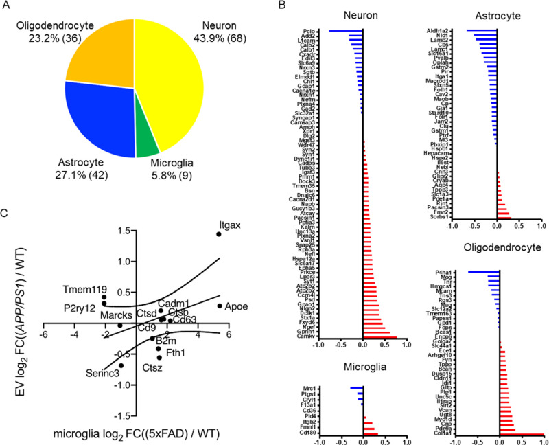
Figure 3.
Cell type-specific protein comparison of CAST.APP/PS1 and WT mouse brain-derived EVs. (A) Enrichment of brain cell type-specific markers in brain-derived EV proteins. Yellow: neuron, green: microglia, blue: astrocytes, orange: oligodendrocytes. The parentheses show the number of identified cell type-specific proteins. (B) Comparison of the cell type-specific protein in CAST.APP/PS1-derived EVs and WT EVs. The red bar shows higher expression in APP/PS1 compared with WT, and the blue bar indicates higher expression in WT compared with APP/PS1. (C) Comparison of log2 fold change of the differential mRNA expression of DAM versus homeostatic microglia in the 5xFAD (x axis) to the log2 fold change of the differential EV protein expression of CAST.APP/PS1 versus WT (y axis).

