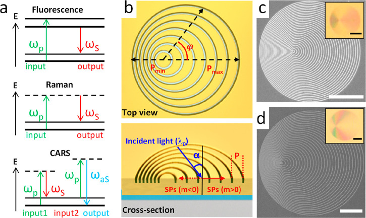
Figure 1.
(a) Schematic illustration of the transitions in the processes of photon-excited fluorescence, Stokes Raman scattering, and two-color CARS. (b) Design of PDG shown in the top view (upper panel) and the tilted view (lower panel). φ is the in-plane azimuthal and α is the incident angle relative to the surface normal. (c, d) Scanning electron microscope (SEM) images of the PDG structures used in this work for (c) SEF/SERS with Δr = 450 nm, d = 140 nm and for (d) SECARS with Δr = 700 nm, d = 400 nm. Their optical reflection images are shown in the insets. All scale bars are 10 μm.

