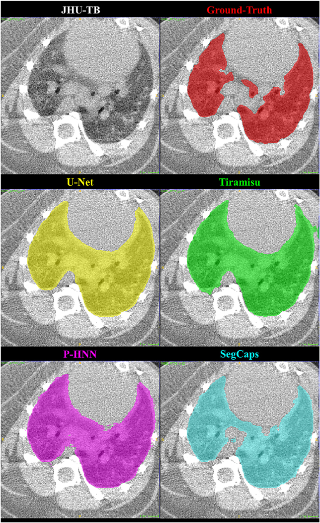
Figure 8:
Qualitative results for a 2D slice from a CT scan taken from the JHU-TB dataset. Note this drastically different anatomy and high level of noise present in the preclinical mice subjects. It can be noticed that the CNN-based methods’ typical failure cases are where the pixel intensities (Hounsfield units) are far from the class mean (i.e. high values within the lung regions or low values outside the lung regions).

