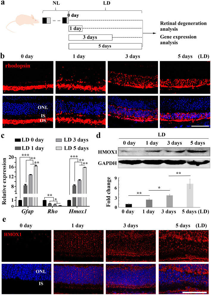
Fig. 1.
The level of HMOX1 induction depends on the extent of retinal degeneration. a Schematic representation of time frame and analysis of light damage (LD). Albino mice were raised in normal light (NL) for 2 months and then exposed to constant white light of 15,000 lx (LD) for 0, 1, 3 or 5 days to induce retinal damage. b Rhodopsin immunostaining of retinas from 2-month-old albino mice kept under high intensity light (15,000 lx) for 0, 1, 3 or 5 days. c Quantification of PCR show the expression levels of Gfap, Rho and Hmox1 in the retinas under the indicated conditions (Error bars: SD; n = 3, one-way ANOVA). d Western blots of anti-HMOX1 and anti-GAPDH in the neural retinas of albino mice after light damage (upper panels) and bar graphs showing quantification of HMOX1 expression according to results of the above blots (lower panel) (Error bars: SD; n = 3, one-way ANOVA). e Anti-HMOX1 immunostaining of frozen sections of retinas from albino mice under the indicated conditions. ONL, outer nuclear layer; IS, photoreceptor inner segments; OS, photoreceptor outer segments. * or ** or *** indicates p < 0.05 or p < 0.01 or p < 0.001. Scale bars: 50 μm

