Fig. 7.
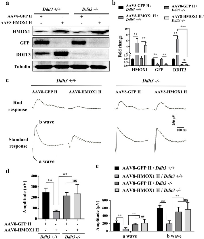
Deletion of Ddit3 prevents abnormal retinal function induced by the AAV8-mediated high dose of HMOX1. a, b Western blots of neural retinas from Ddit3+/+ (n = 3) and Ddit3−/−mice (n = 3) 2 weeks after infection with a high dose of AAV8-GPF or AAV8-HMOX1, using anti-HMOX1, anti-GFP or anti-DDIT3 antibodies (a) and corresponding quantitative bar graphs (b) (Error bars: SD; one-way ANOVA; *, p < 0.05; **, p < 0.01; ***p < 0.001). c ERG traces of 2-month-old Ddit3+/+ (left panels) and Ddit3−/− (right panels) mice 2 weeks after infection with the high dose of AAV8-GFP or AAV8-HMOX1. d, e Quantification of ERG amplitudes in rod response (d) and standard response (e) according to the ERG trace results from c (Error bars: SD; n = 4, one-way ANOVA; *, p < 0.05; **, p < 0.01)

