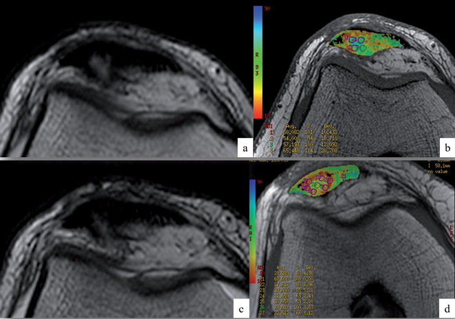
Figure 1.

Axial images of standard T2 (a, c) and T2 mapping (b, d) sequences in a patellar tendon, before (a, b) and six months after (c, d) percutaneous US-guided intratendinous PRP injection treatment. Note the reduction of mean T2 relaxation values of circular ROIs within the most tendinopathic area (visible as a hyperintense intrasubstance alteration on standard imaging) after treatment, consistent with signs of tendon healing
