
Figure 2.
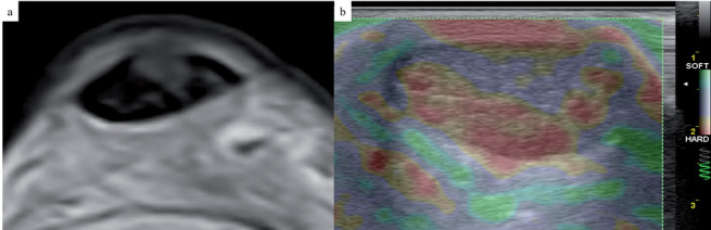
Axial T2-weighted image (a) and short-axis compression US elastography image (b) of an Achilles tendon. Note the intrasubstance hyperintense area of tendinosis within the tendon, evident at a “softer” area (in purple) at US compression elastogram