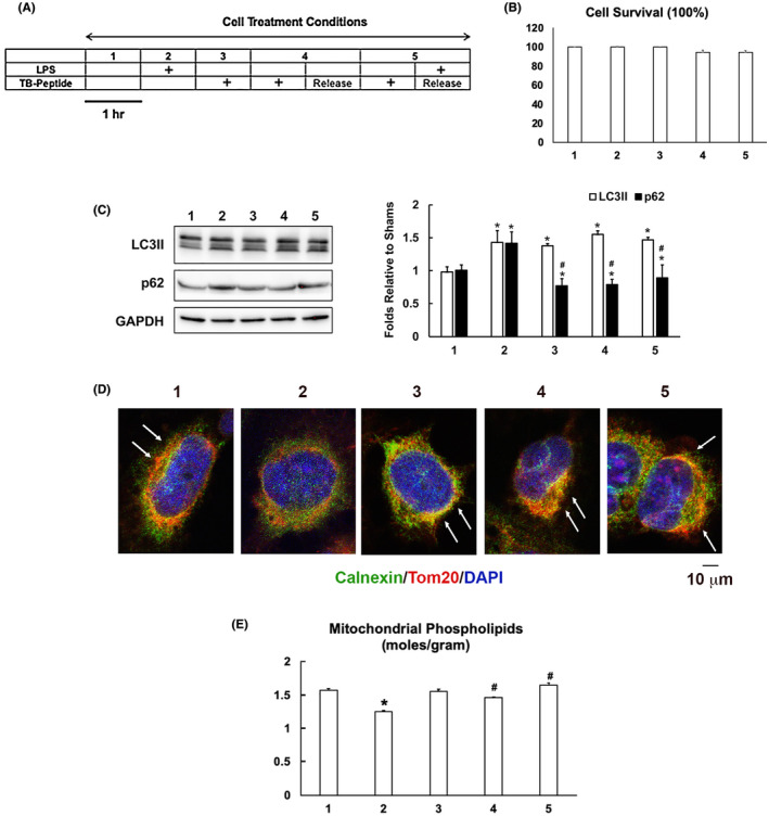
FIGURE 5.

TB‐peptide promotes autophagy and protects MAMs in human cardiomyocyte challenged by LPS. A, AC16 cells were cultured until 80%‐90% confluency and treated with the conditions as illustrated. B, The percentage of cell survival was calculated based on the analysis of cytotoxicity. C, Levels of LC3II and p62 in cell lysates were examined by western blot and quantified by densitometry. GAPDH was used as a loading control. D, Cells were co‐immune‐stained with the ER marker calnexin (green) and mitochondria marker Tom 20 (red). Staining of nucleus by DAPI is shown in blue. Overlay areas of mitochondria–ER contact shown in the color yellow indicates the levels of MAMs and are labeled with white arrows. All images are representative of ≥3 independent experiments. E, Levels of phospholipids in mitochondrial fractions were quantified and results were normalized by the amount of protein. All values are means ±SEM. Significant differences are shown as * for sham vs LPS‐treated and # for without vs with TB‐peptide (p < .05, n = 3‐5, unpaired t test)
