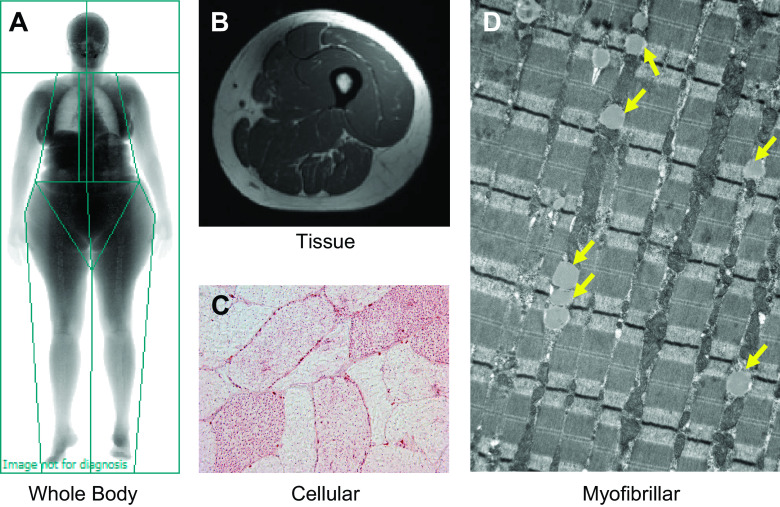
Figure 1.
Different measurement techniques can be used to quantify adiposity deposited at various anatomical locations. Briefly, adiposity can be assessed at the whole body (A), tissue (B), cellular (C), and subcellular (D) levels. At the whole body level (A), dual-energy X-ray absorptiometry scans yield measurements of overall adiposity (e.g., percent body fat) and regional estimates (android vs. gynoid deposition). Magnetic resonance (MR) imaging and computed tomography (not shown) scans can be used to characterize specific fat depots (B), such as visceral adipose tissue surrounding the organs of the abdomen and intermuscular adipose tissue of the thigh. Superconducting magnets also offer the capacity to examine the chemical composition of tissues, and MR spectroscopy can be used to provide a noninvasive estimate of intramyocellular lipid content. Finally, the number, size, and area of individual lipid droplets can be measured at the cellular (single fiber) level (C) using oil-red-o immunohistochemistry as well as at the myofibrillar level (D) with electron microscopy. Yellow arrows in electron micrograph indicate the presence of individual lipid droplets.

