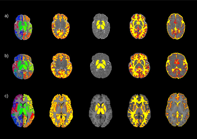
Figure 1.

An example of the parcellation and the segmentation from three different subjects: a) a preterm born baby, b) a term born baby, and c) an adult subject. From left to right, the parcellation and the four different tissue probability maps included in the five tissue type file: gray matter, subcortical gray matter, white matter, and cerebrospinal fluid. For the neonates, the maps are overlaid onto the T2w volumes for the neonates and onto the T1w volume for the adult.
